

Radio Vision - Scientific Milestone
A Research Activity of Drown Laboratories, 1960

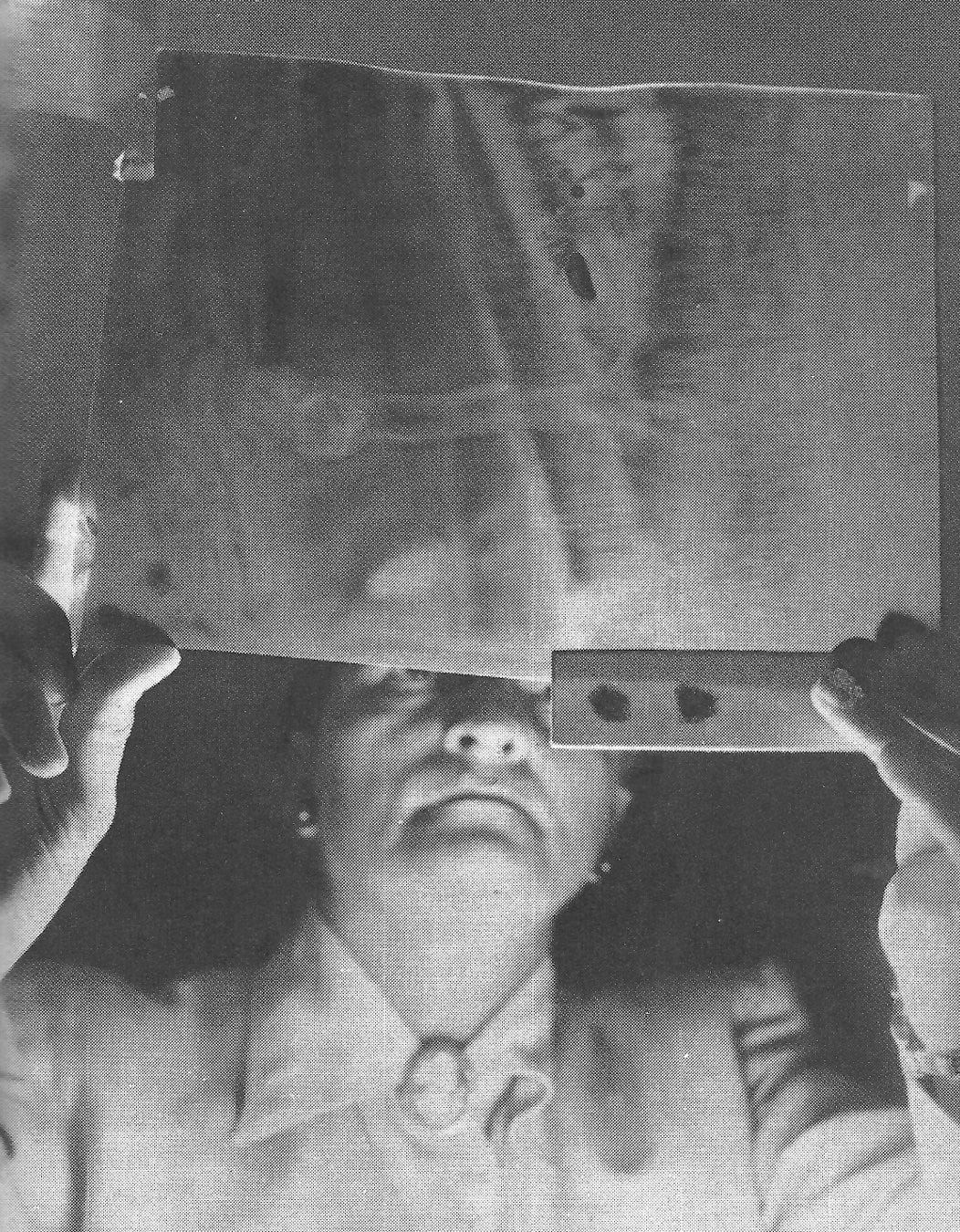
In the mid-20th century, the work of Dr. Ruth B. Drown and her groundbreaking invention, the Drown Radio-Vision Instrument, represented a bold and controversial chapter in the history of visionary medical technologies. This booklet, originally published in 1960 by the Humanitarian Research Foundation in association with Drown Laboratories, serves as a historical artifact showcasing the purported capabilities of Radio-Vision—a method claimed to capture cross-sectional photographs of human tissues using only a patient’s “blood crystal.” These pictures were taken in total darkness, with no visible light on the instrument.
While the scientific community largely dismissed these claims, the instrument’s ability to transcend geographical boundaries and provide diagnostic insights captivated proponents of unconventional medical approaches. Behind the scenes, this publication was quietly supported by Trevor James Constable, well known for his pioneering explorations into subtle energies and etheric phenomena. Presented here as a historical reprint, which I scanned from a copy Trevor gave me long ago to preserve, this document offers a glimpse into a fascinating yet contentious attempt to merge technology, biology, and metaphysics in pursuit of revolutionary medical diagnostics. It is reproduced verbatim, without inserted comment, to preserve the record accurately.

RADIO-VISION Scientific Milestone
A RESEARCH ACTIVITY OF DROWN LABORATORIES
Sustained by the HUMANITARIAN RESEARCH FOUNDATION
© Dr. Ruth B. Drown 1960
The Drown Radio-Vision instrument was invented in 1935 by Dr. Ruth B. Drown of Los Angeles, California, U.S.A. Radio-Vision is the only known method by which pathological and histological cross sectional photographs of the soft tissue and hard tissue of the human body may be obtained. Radio-Vision photographs are made regardless of the geographical separation between the Instrument and the patient.
The Radio-Vision instrument provides access to areas and organs of the human body inaccessible for examination other than by surgery. Radio-Vision photographs enable the trained physician to make careful studies of the pathological conditions prevailing in the organs, glands and tissues of the human body. The photographs are made from the blood crystal of the patient, held in a chip of blotting paper. The energy pattern of the human body passes through this blood crystal as long as the patient lives. This energy is used to make the photographs.
The theory on which Radio-Vision is based is extremely simple. It may be considered so simple that it has been overlooked and ignored in the pursuit of more ramified techniques, none of which has yet yielded the vivid results provided by Radio-Vision.
Fundamentally, the theory is based on the fact that everything having form in the physical world is made up of molecules. The molecular arrangement establishes the outer form of the substance. Because the molecular arrangements producing liver tissue for example, are different to the molecular arrangements producing lung tissue, liver tissue and lung tissue differ from each other in their outer form.
The molecules consist of whirling particles of electricity. This motion produces a definite emanation from all physical substances, which may be brought under direct observation through the specialized use of pinacyanole bromide filters and screens.
Differing molecular arrangements producing differing forms, must also produce differing characteristic emanations in each case. In general terms, they produce differing frequencies or vibrations.
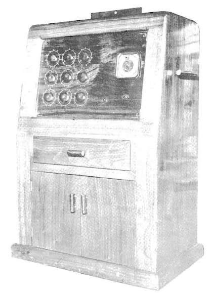
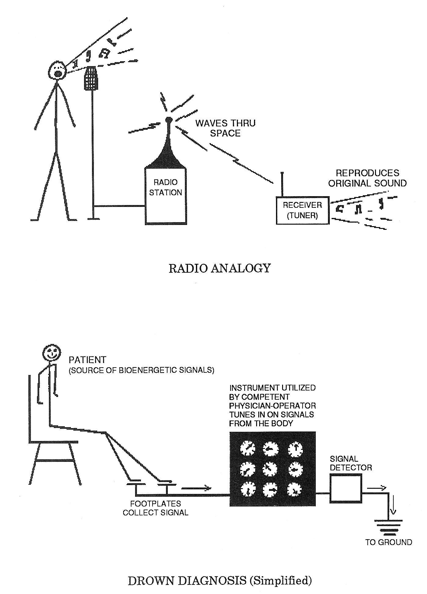
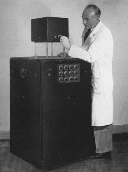
These emanations may be detected and numerically classified on the Drown Diagnostic Instrument, also invented by Dr. Ruth Drown. The Drown Diagnostic instrument is a very simple impedance rheostat, consisting of nine dials, each of which can select ten tuning stubs by its rotation. Each dial is numbered from 1 to 10, each dial position making contact with a stub. The possible combinations permitted by this arrangement exceed two billion.
Systematic use of the Drown Diagnostic Instrument through the past thirty years has established that healthy human tissues and organs have definite “Rates” of vibration, or molecular broadcast frequencies, which the diagnostic instrument can detect. The entire human body has been assigned “Rates” as they are termed, for all organs, glands and tissues.
Diseases have also been found to have characteristic rates. Pathological tissue, for example, no longer has its original form. The changes in form produce changes in the emanations of the molecules. All the major diseases and many minor ones have been systematically assigned their rates and these have been found to be the same throughout the entire human family,
These processes amount to the expression of healthy tissues or organs as numbers and diseases as numbers. This has permitted the development of a very exact and remarkable system of diagnosis. In the hands of a competent and knowledgeable physician, trained in its use, the Drown Diagnostic instrument offers the possibility of unerring diagnosis. The findings of the Instrument verify that the foundations of the Universe are mathematical.
In the course of the development of this system of diagnosis and therapy, it has been established firmly that a blood crystal from a human being, held in a piece of blotting paper, carries the complete energy pattern of the owner’s body. The blood crystal is perpetually resonant to the owner, no matter where on earth the owner may be. From the blood crystal the same diagnosis may be made as when the patient is present in the doctor's office. The Radio-Vision photographs are made the same way.
The blood crystal is the visible end of an invisible line connected to the owner as long as he lives, and for a short time afterwards.
Radio-Vision is used in conjunction with the Drown diagnosis. The diagnosis establishes the presence of certain conditions of disease or malfunction in the patient. The tuning section of the Radio-Vision Instrument is set to the rate of the condition it is desired to photograph. The film, ordinary panchromatic material size 8X10", is inserted in the slot, removed a few seconds later, and processed in ordinary chemicals.
The photograph that appears is a histological or pathological cross section of the area into which the Radio-Vision Instrument has been TUNED. The conditions existing in the patient must have been established by diagnosis, the rates of those conditions found before the Radio-Vision Instrument can be used. The Radio-Vision Instrument is therefore not a machine that can be operated by push-buttons, or by incompetent and ignorant persons. It is an instrument of superlative sensitivity intended for use by a thoroughly trained, competent and wise physician.
If at times the structures do not appear exactly as the physician might expect, this may be due to uneven response of the film to these subtle energies. Much work has yet to be done on the photographic materials involved, and their relationship to the energy they are recording.
The photographs have been made through the blood crystals of the patients involved, except for two of the human brain, which are appropriately captioned. Distances separating patients from the Instrument varied from one floorof a building to eleven thousand miles. The Atlantic Ocean or a continent is spanned as easily as a city block by this new mutation of energy, connected with the life of human beings, and manipulated by the Drown Instruments.
A basic interpretation of each photograph has been given, and arrows placed on the photographs to orient the reader. The physician will have no trouble in following the amazing record herein set forth, and nor will the intelligent layman.
What the physician will surely recognize is that in these photographs, some of them made before panels of eminent medical men, one of the great dreams of medical science has come to fruition. Soft tissue cross-sectional photographs are a reality, and have been for twentyfive years. The responsibility rests with the medical profession to see that this work is taken up on the broadest possible basis, so that its benefits may flow out to humanity.
Through the years, many of the Radio-Vision pictures have been verified by post-mortem surgery. In what they show, in what they tell the learned medical man, they are their own proof of the efficacy of the technology that produced them.
Scientists outside the medical field must recognize the vast potential that this technology holds. In the fields of mineral prospecting, space technology, agriculture, nuclear physics, special applications glitter before mankind. And they are just a few.
New research is carrying this work to a still higher plane of achievement, with no end in sight to what may be recorded by these methods. New designs in the instruments, moving constantly towards even simpler arrangements are revealing ways to tap into cosmic laws and secrets never hitherto accessible to man. Special sensitized materials and devices open incredible new vistas for the human race.
The lesson of these photographs, and of the work of Dr. Ruth B. Drown, inventor of the Radio-Vision Instrument and of Drown diagnosis and therapy, is this;
“Everything is here, now. All we have to do is tune in on it.”
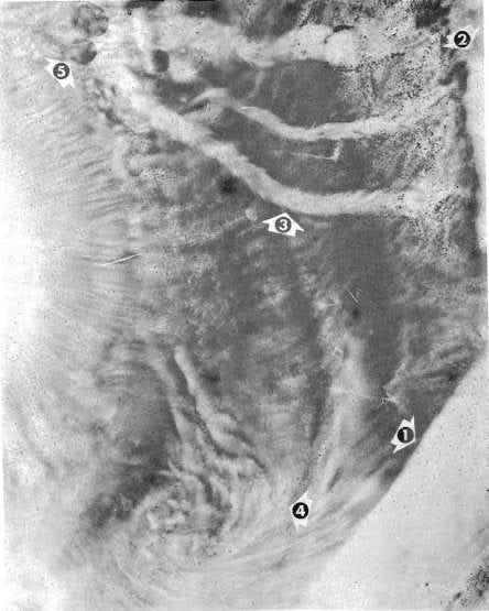
PLATE No. 1
The Sacral Plexus Region
Arrow 1 shows a cross section of the sacral bone. Arrow 2 shows portions of the vertebrae. It is as though the patient were facing the left side of the picture, and his pelvic region vertically cross sectioned by a gigantic knife. Arrow 3 shows three blood vessels in a state of diapedesis, carrying blood to the meningeal coccus infection concentrated at the ends of the blood vessels near the vertebrae. The meningeal coccus, on which the Radio-Vision Instrument was tuned, shows as myriad black dots all through the region photographed. The infection shows in the bottom right hand corner, in the sacral bone itself; it shows through the structure of the gluteal muscle (Arrow 4). The black circles, (Arrow 5) are blood vessels which travel at right angles to the plane of the picture.
The infection was detected first by the Drown Diagnostic Instrument. Then the Radio-Vision Instrument was tuned in on the infection, using the rate established in the diagnostic process. The patient was seven city blocks away from the instrument when the picture was made.
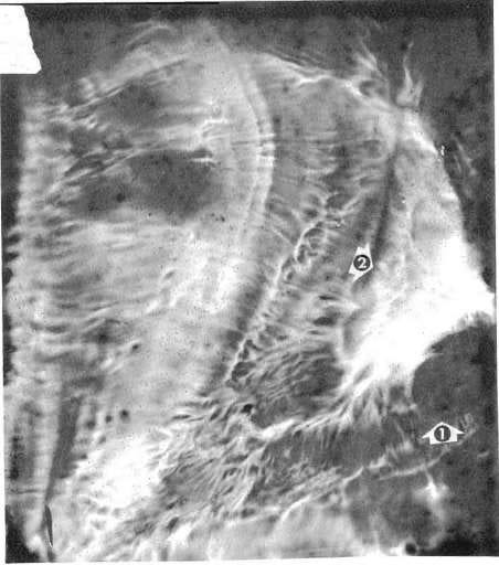
PLATE No. 2
Gallstone in Gall Bladder
This Radio-Vision photograph was made by tuning in on the gallstone, detected in the Drown diagnosis. The photograph is a perfect cross section of the gallbladder, the stone itself appearing imbedded in the tissue (Arrow 1). The inflammation (arrow 2) shows above the stone itself. The almost three dimensional nature of the picture shows the startling clarity with which these soft tissue pictures may be made. The patient was separated from the instrument by one floor of a building and connected to it only by her blood crystal.
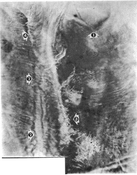
PLATE No. 3
This Radio-Vision photograph shows the Thyroid Gland (arrow A) in a case of Hodgkins disease. The two arrows (2) show the spinal cord, deleting the vertebral bony structure. The numerous black circles indicated by Arrow 3 are cross sections of blood vessels in the pleura of the lungs. Arrow 4 indicates the pleura.
The patient was 1300 miles away when the picture was made via the blood crystal, Drown diagnosis having previously verified the presence of Hodgkins’ disease.
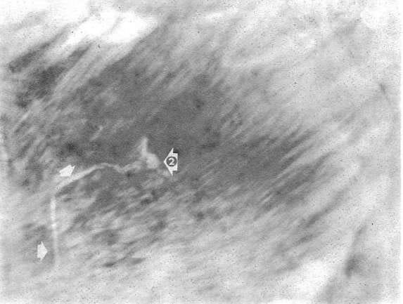
PLATE No. 4
Pathway of Bullet in Patient’s Chest
A domestic servant of Dr. Drown in the years before World War II feared that her son had been injured by a rifle shot, aimed at him indiscriminately by young hoodlums passing in a car. Knowing of Dr. Drown’s photographic instrument, she brought a crystal of the boy’s blood to Dr. Drown.
Dr. Drown took two photographs by Radio-Vision. The first photograph, this one, saw the Radio-Vision Instrument set to the injury or wound rate. The arrows indicate the pathway through the tissue of the chest followed by the bullet, which had in fact lodged in the boy’s body. Arrow 2 indicates the area of final lodgement of the projectile.
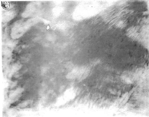
PLATE No. 5
Actual Bullet in Patient’s Chest
This Radio-Vision photograph was made immediately following Plate 4. The tuning of the Radio-Vision Instrument was altered to tune in directly on the bullet itself, rather than on the wound. The arrow shows the actual bullet end-on, and it measures on the original 8 X 10 photograph exactly .22 inches.
The boy with the bullet in his chest was 22 miles away at the time this Radio-Vision photograph was made, the only connection with the Radio-Vision Instrument being his blood crystal, supplied by his mother.
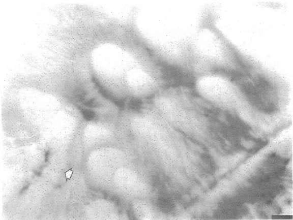
PLATE No. 6
Ovum in an Ovary
This Radio-Vision photograph shows Oophorons in different phases of development in an ovary. The vivid, egg-shapes are undeniable. Arrow shows a ruptured oophoron in the lower left corner of the picture.
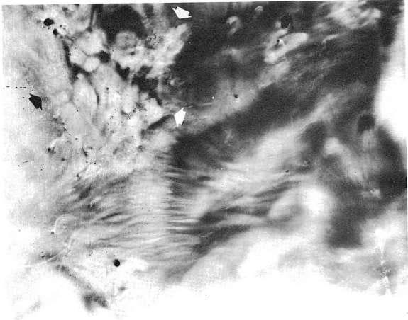
PLATE No. 7
Cancer of the Stomach
This picture was thought by a number of doctors to be a diaphragmatic hemia. However, the Drown diagnosis recorded cancer of the area shown, and it was into this that the Radio-Vision Instrument was tuned. The arrows show the outline of the cancer. Numerous blood vessels show as black spots and spheres, as they run at right angles to the plane of the photograph.
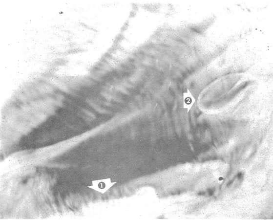
PLATE No. 8
Ulcer of the Stomach and Scirrhous Cancer
This Radio-Vision photograph is a cross sectional picture of the stomach of a patient whose diagnosis showed both scirrhous cancer and an ulcer. The cancer shows as the white, thickened tissue (Arrow 1), the ulcer on the right being clearly outlined (Arrow 2). Photograph made from patient’s blood crystal, with patient remote geographically from the Radio-Vision Instrument.
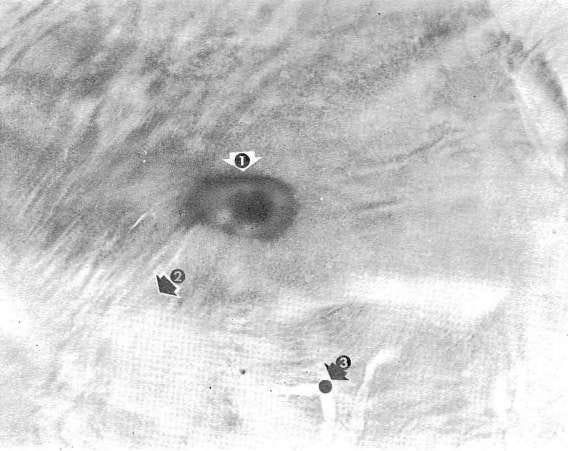
PLATE No. 9
Bony Structure of the Head
The Radio-Vision Instrument was tuned in on the bony structure of the head for this photograph. The actual structure of the bone will be readily recognized by physicians. Arrow 1 shows the auricular area (ear), (Arrow 2) the cerebellum area, and (Arrow 3) a blood vessel cross sectioned by the photograph. The patient would be looking towards the right hand edge of the photograph.
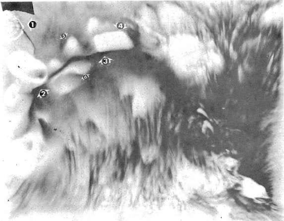
PLATE No. 10
Surgical Scar and Type 4 Cancer (Breast Area)
This patient was in Illinois, the photograph being made in Los Angeles via blood crystal. Arrow 1 shows the nipple to orient the reader. The black line (Arrows 2 and 3) shows the scar of previous surgery to remove the cancer. The white areas around the scar, (Arrows 4, 5, and 6) show how the cancer has returned.
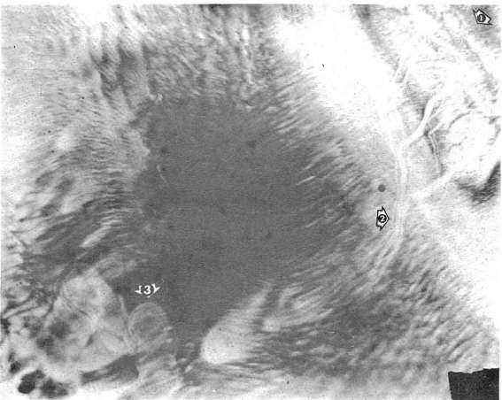
PLATE No. 11
Cancer of Portion of Colon
Drown diagnosis verified the presence of cancer in this patient’s colon. Arrow 1 shows the cancerous mass in the colon. Arrow 2 shows blood vessels extending from the cancerous area. Arrow 3 shows a large blood vessel cross sectioned by the Radio-Vision Instrument.
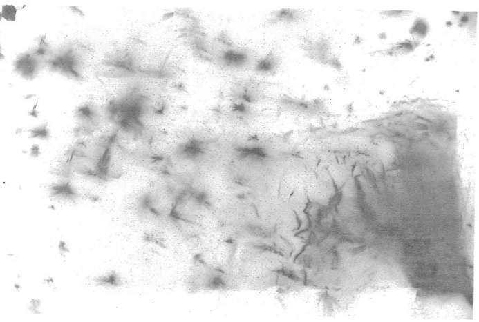
PLATE No. 12
Energy in the Brain (Awake)
This photograph, and plate 13 which follows, were among the first Radio-Vision photographs made. These were obtained by placing the patient’s head between 2 collector pads, a technique that became obsolete when the pattern-bearing properties of the blood became established. One of the first research steps in Radio-Vision, this photograph was made of a young woman in California when she was awake. The electrical activity of the brain is strongly indicated by the patterns recorded.
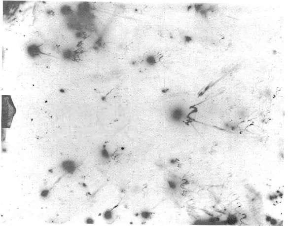
PLATE No. 13
Energy in the Sleeping Brain
When the young woman dropped off to sleep, Dr. Drown made this photograph, showing a distinct contrast from the waking brain in Plate No. 12. Certain of the forms repeat themselves many times. These photographs (Plates 12 and 13) were exhibited at the Iowa State Fair and published in “Look” magazine in 1937.
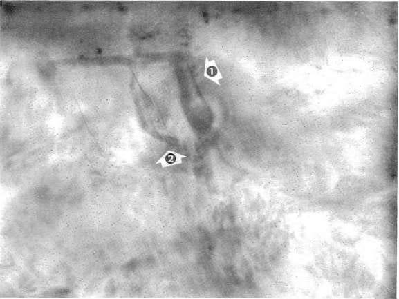
PLATE No. 14
Surgical Instruments Photographed Across the Atlantic
The patient, an old English friend of Dr. Drown’s, wrote her in 1950 that he was to have an operation for an ischio-rectal abscess. He asked Dr. Drown to take a Radio-Vision picture before the operation, due the day following Dr. Drown’s receipt of his letter. At 1 a.m. the following morning Dr. Drown tuned in on the man’s abscess and this picture resulted. Arrow 1 shows the surgeon’s retractors. Arrow 2 shows a pair of scissors or a hemostat, in the immediate region of the abscess. Subsequent comparison of times showed that the patient was under surgery at the time photographed.
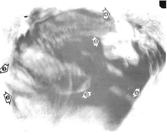
PLATE No. 15
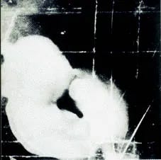
Human Foetus at Approximately Three Months
This Radio-Vision photograph was made from the blood crystal of the mother. The foetus, although physically very small at the time, has filled this picture with its head, an enormous magnification. Arrow 1 shows the chin and mouth area. Arrow 2 shows the nose, to the right of which the forming eye and forehead may be seen. Arrow 3 shows the forming ear of the child. Arrow 4 shows the energy entering the head of the embryo through the top of the head. Arrow 5 shows this vital energy in the region of the pineal gland, settling an ages-old controversy. Arrow 6 shows a portion of the yolk sac.
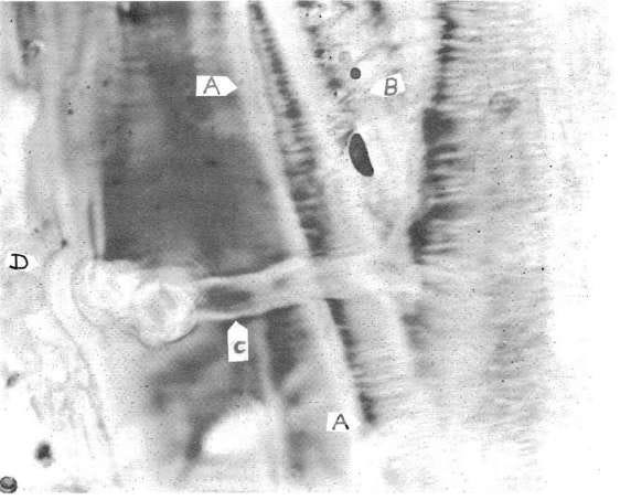
PLATE No. 16
Abcessed Tooth
This photograph was made by tuning in on the abscess. The outline of the root of the tooth is shown by the arrows A. Arrow B shows the pulp of the tooth, with the black and dark spots blood vessels within that pulp that have been cross sectioned by the Radio-Vision Instrument. Arrow C shows a sinus of infection from the lower part of the pulp area through the gum of the patient. Arrow D shows the gum boil as a bulge in the mouth of the patient.
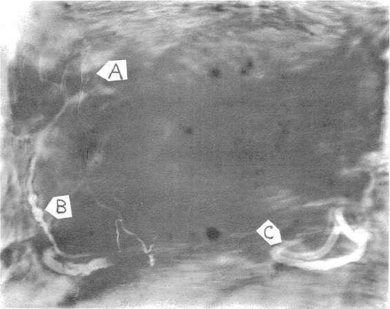
PLATE No. 17
Coronary Occlusion of the Heart
This picture was made after death. The heart valves are indicated by Arrow A. The occlusion is shown by Arrow B, which points to the clot causing the heart failure. Other blood vessels in the heart are shown by Arrow C. The photograph may be compared with anatomical drawings of the heart which show the same areas and detail.
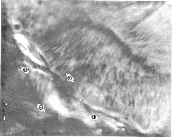
PLATE No. 18
Tumor of Eustachian Tube of Ear
This patient complained of increasing deafness. A Drown diagnosis showed a tumor to have formed in the region of the Eustachian tube or auditory canal. The Radio-Vision instrument was tuned in on this tumor. The line of the Eustachian tube is shown by Arrows 1. The base of the tumor is shown by Arrow 2. The upper portion of the tumor (3) has compressed the Eustachian tube, causing the deafness of which the patient complained.
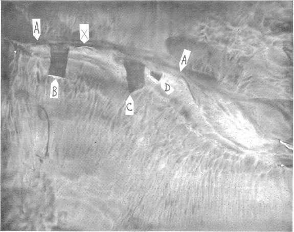
PLATE No. 19
Post Surgical Picture - Abdomen
The line of the incision is indicated by the Arrows A. B, C and X are blood vessels severed during the operation. The whitish effect below the line of the incision is due to inflammation. Arrow D shows a smaller blood vessel, also severed.
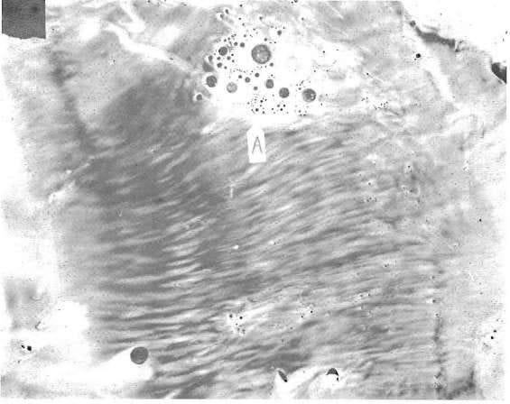
PLATE No. 20
Hydatid Cyst in Liver
Cross section of patient's liver, made through a blood crystal. The patient recorded a hydatid cyst or mole in diagnosis by Dr. Drown. The Radio-Vision Instrument was tuned in on the cyst, shown by Arrow A. The numerous other black circles and dots are bile ducts and blood vessels. The tissue structure should be compared with that shown in Plate 21, also a liver photograph.
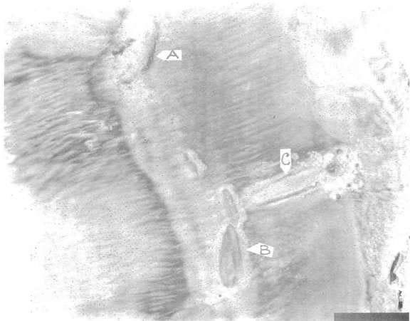
PLATE No. 21
Cancer Craters in the Liver
Arrows A and B show cancer craters in the liver of a patient residing in Indiana, the photograph being made in Hollywood, California from her blood crystal. The similarity of the liver tissue to that shown in Plate 20 should be noted. Arrow C shows a needle lesion in the patient's liver from prior treatment.
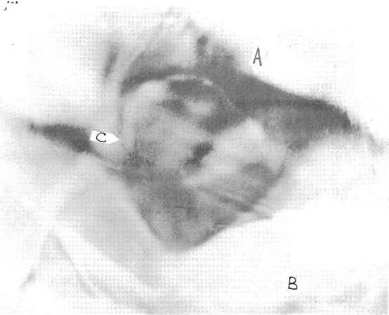
PLATE No. 22
Blood Clot and Cancer at Pyloric End of Stomach
This Radio-Vision photograph was made in England in 1939 by Dr. Ruth Drown, before an audience of seven British medical doctors, As she was discussing her work and methods, the patient’s blood crystal arrived from America, having originally been sent to her Holly wood office by Dr. Henry Pratt, a Connecticut osteopath. Symptoms included the patient’s inability to ingest or pass his food. Dr. Drown diagnosed the patient and stated that the man had cancer of the stomach and a blood clot recorded also. Tuning in on the blood clot, Dr. Drown made this picture in front of the M.D.s. Arrow A shows the line of the pyloric end of the stomach, which extends from the right hand edge of the picture. Arrow B shows the white, thickened, cancerous tissue. Across the funnel-like opening of the stomach between A and B is the blood clot, C. The picture was sent out the same night on the trans-Atlantic clipper. Dr. Pratt received it shortly before the patient died. Post mortem surgery revealed the clot and the cancer to be exactly as shown in this photograph, taken from London, England, the previous night.
CONCLUSION
You have had your introduction to what is in all probability the greatest single invention of our time. You have seen with your own eyes photographs of the internal organs of the human body. You have seen photographs of various pathologies and foreign bodies. You have been presented with the evidence of the manner in which this instrument annihilates time and space.
The technology behind the Radio-Vision instrument exists as a successful system of diagnosis and therapy, it has been used and verified and welded into a useful system since Dr. Drown’s first invention in 1929. The Radio-Vision instrument has been in existence for twentyfive years, largely uncomprehended in its significance and capacity to serve mankind.
To the reader who responds with “Has this been proven?” his attention is redirected to the photographs. There is the proof! There is no other way of making such photographs, and no way of “verifying” them, other than through the use of an inferior method.
Radio-Vision is a great diagnostic tool, a great investigative tool in other fields, a contribution to civilization and mankind's march towards the light.
This booklet has been produced to acquaint physicians and intelligent laymen alike with the properties and performance of this Instrument. Many illusions exist concerning this work, which this book will help remove.
Radio-Vision is indeed a scientific milestone.
Sustained by THE HUMANITARIAN RESEARCH FOUNDATION
****** finis ******
After thoughts:
Trevor James Constable, in his seminal work The Cosmic Pulse of Life, regarded Ruth B. Drown as a visionary whose work transcended the boundaries of conventional science and delved into the esoteric realms of life energy and cosmic forces. He admired her pioneering approach to radionics, particularly her belief in the subtle, life-related energies that underpin existence—an idea that resonated with his own explorations into etheric phenomena. He connected the vital energy of her radionic work with that of Wilhelm Reich’s orgone energy.
Constable saw Drown’s Radio-Vision Instrument as a profound attempt to bridge the gap between physical and metaphysical realities, aligning with his broader view that humanity is deeply interconnected with unseen cosmic energies.
In Cosmic Pulse, Trevor noted the parallel development in the work of George and Marjorie de la Warr, whose radionic camera shared conceptual similarities with Drown’s invention. The de la Warr camera purportedly captured images of the past and future, further illustrating the potential of these devices to access dimensions beyond ordinary perception. The de la Warr camera was apparently unknown at the time of the publication of Radio-Vision: Scientific Milestone, as it speaks of “no other” similar advances.
Together, these innovations reflect a bold epoch in alternative science, where visionaries like Drown and the de la Warrs sought to unveil the hidden patterns of life and consciousness, challenging the materialist paradigms of their time.

Support Independent Research into Hidden Knowledge & Unexplored Dimensions
Thank you for reading, I hope you enjoyed this. Your engagement enables deep investigation into fundamental aspects of existence that mainstream academia often neglects. This work examines critical historical and scientific understanding through alternative perspectives.
🎯 Make a Direct Impact:
PayPal: paypal.me/alkemix33
Purchase a course: https://alkemix.gumroad.com/
Why Support?
Each contribution facilitates:
Investigation of overlooked historical evidence
Research into alternative scientific paradigms
Documentation of significant findings
Creation of original content that challenges conventional narratives
Whether you:
Share these insights with fellow researchers
Contribute to ongoing investigations
Engage in meaningful discourse
You're helping illuminate paths of knowledge that deserve deeper examination.
"The gift is to the giver, and comes back most to them" - Walt Whitman
What people say about my work:
“Thomas’ lectures are worth watching several times. Gold in every minute. So grateful for them.” —JE
“The absolute best. It always fills me with joy to watch Thomas’ lectures, restores my sanity in these crazy times.” —KS
“I so love your work. You’re a beautiful human angel.” —JG
“Your work is very important, continues to be a solid support and inspiration to me through all the madness, Thomas - thank you!” —K
“Thomas Brown has labored long and mightily in that most difficult of all assignments: presentation of new ideas and conceptions to a world determined to go to hell.” —Trevor James Constable
I was just at the Wilhelm Reich museum, Orgone, in Rangeley Maine, and there is a wealth of overlooked and misunderstood technology that needs to be brought back out by competent researchers.
In the borderland research museum is an absolute treasure trove that I fear has been relegated to oblivion.
Perhaps NOT... And here is some rather striking evidence...
https://quackwatch.org/chiropractic/hx/drown/